Diagnosing Myositis
Diagnosing Myositis can be a long and frustrating journey. It is not unusual to be seen by several doctors over months or years. Unfortunately, it is also not unusual to be misdiagnosed, thereby delaying effective treatment or being given the wrong treatment. Some reasons this may happen are:
Types of Physicians who diagnose and treat Myositis
Keep in mind that the individual types of Inflammatory Myopathies are rare diseases that include a variety of symptoms such as muscle weakness, muscle pain, trouble swallowing, skin rashes, breathing difficulties, fever, fatigue, joint pain and swelling, vocal changes, etc. Some myositis patients have many of these symptoms while others may have only a few.
During your journey towards a diagnosis of Myositis, you will likely visit many specialists depending on your symptoms. Some of the doctors you may see include a Rheumatologist, Neurologist, Neuromuscular specialist, Dermatologist, Pulmonologist, Cardiologist, Oncologist, Gastroenterologist, pain management, and others depending upon which organs are involved. These will likely be coordinated by your Primary Care Physician.
What to expect during the diagnosis phase:
Medical and Family History and Clinical Exam
An office visit with a physician will likely include asking about a patient’s medical history and that of their family members. Some doctors feel that a patient’s medical and family history is one of the most important parts of helping to diagnose an illness, especially a complicated one like Myositis. Before your appointment, take the time to write down any important medical history and share this with your doctor.
The visit should include a physical examination to test to assist in the diagnosis and may include:
Diagnostic Testing for Myositis
In addition to a clinical examination and discussion of existing symptoms and history, physicians often conduct many diagnostic tests to assist in narrowing down a diagnosis. Tests include markers for autoimmunity, inflammation, and autoantibodies.
Blood Tests
Many patients with Myositis have elevated levels of muscle enzymes which may indicate the presence of muscle damage. Enzymes act as catalysts for chemical reactions that take place in cells. Many enzymes are normally present in the blood, but when cells are damaged by disease or injury, large amounts leak out, which can cause elevated levels. Keep in mind that there are also many patients who do not present with the typical elevated muscle enzymes.
Aldolase is a protein (enzyme) that helps break down certain sugars to produce energy. It is found in high amount in muscle tissue. Tests are conducted to diagnose or monitor muscle damage, and elevated Aldolase levels can indicate damage to the muscles.
ALAT/ALT/SGPT (Alanine aminotransferase, serum glutamate pyruvate transaminase), is an enzyme found in muscle, heart and liver tissue. ALT is not used as a stand-alone diagnostic test.
ASAT/AST/SGOT (Aspartate aminotransferase, serum glutamic-oxaloacetic transaminase), is an enzyme found in muscle, heart and liver tissue. AST is not used as a stand-alone diagnostic test but can provide some indicators your doctor may wish to monitor.
Creatine Phosphokinase (CPK), also referred to as Creatine Kinase (CK), is an enzyme found mainly in the heart, brain, and skeletal muscle. When muscle is damaged, CPK/CK leaks into the bloodstream and an elevated level usually means there has been injury or stress to muscle tissue which are indicators for polymyositis, dermatomyositis, other myopathies and muscular dystrophies. Some patients with a confirmed diagnosis of PM or DM may not have elevated CK levels, even during active disease.
Normal range for CPK/CK is approximately 22 to 198 IU/L (units per liter), however, the range can vary depending on gender, the laboratory used, and what your physician considers “normal.”
Note: The CK or CPK values can also indicate damage to the heart, brain or lungs, but specific CK/CPK tests would be ordered by your doctor to make this determination.
Factor VIII-related antigen (also called von Willebrand factor VIII-related antigen) shows damage to the lining of blood vessels and can help physicians see the extent of the problem, especially in Juvenile Myositis (JM). This may assist in determining a treatment plan.
Flow Cytometry looks at a specific group of white blood cells and is often used to learn more about the extent and severity of Juvenile Myositis (JM).
LDH (Lactate dehydrogenase) is an enzyme found in skeletal muscle tissue. High levels in the blood can be a general indicator of tissue and cellular damage or show the existence and severity of acute or chronic tissue damage but can provide some indicators your doctor may wish to monitor. (Methotrexate, one of the treatments used for myositis, may cause an elevated LD level)
Sed Rate (also called ESR or Erythrocyte sedimentation rate). This test can reveal inflammatory activity in the body. It is not a stand-alone test but may help with diagnosis and monitor a patient’s progress.
For sIBM: There is blood test available, since 2013, specifically for IBM. Ask your physician.
Do you have normal blood work?
If you are like some other patients with a now confirmed autoimmune myositis diagnosis and you have a normal CK and/or aldolase level, Dr. Andrew Mammen explains:
For CK to leak into the bloodstream there needs to be some muscle necrosis and with dermatomyositis that may not be found. Listen to Dr. Mammen in this podcast.
Antibody Tests
ANA (Antinuclear Antibodies panel) The immune system normally makes antibodies that help fight infection. Antinuclear antibodies often attack the body’s own tissues (autoimmune). In this case, these attack a cell’s nucleus. This testing does not confirm a specific disease diagnosis, however specific patterns can assist with a differential diagnosis.
A common misunderstanding among patients is that the ANA is only used to test for Lupus.
Myositis-Specific Antibodies (MSA) MSA’s are almost never found outside of patients with myositis, meaning the presence of these strongly support evidence for a diagnosis. There is a lot of research being done with myositis antibodies.
Myositis-Associated Antibodies (MAA) Myositis-Associated antibodies can be found in patients outside of a myositis diagnosis, however positive test results can provide supporting evidence of association with other diseases. There is a lot of research being done with myositis antibodies.
It is estimated that around 50% of myositis patients have myositis-specific and/or myositis-associated antibodies.
More advanced testing
EMG (Electromyography)
An EMG is performed to identify the location and type of inflammation due to damaged muscle tissue. A thin needle electrode is inserted through the skin into muscles. Electrical activity is measured and recorded as muscles are tensed and relaxed. The doctor or tech performing the EMG will move the needle electrode various times to measure different areas of the same muscle or various muscle locations. Changes in the pattern of electrical activity can confirm muscle disease and distribution.
Nerve Conduction Study (NCS)
A nerve conduction study measures how well and how fast nerves send electrical signals. It evaluates the functional ability of electrical conduction of the body’s motor and sensory nerves. An NCS is used to identify damage in the peripheral nervous system (nerves that lead away from the brain and spinal cord and connect the central nervous system to the limbs and organs). Several electrodes are attached to the patient’s skin, and a shock-emitting electrode is placed over a nerve while a recording electrode is placed over the muscles controlled by that nerve. Electrical pulses are given to the nerve, and the time it takes for the muscle to contract in response to the electrical pulse is recorded. The speed of this response is the conduction velocity. Nerve conduction studies are performed prior to an EMG and may include both sides of the body for comparison. Unless neuropathy exists, someone with Myositis should have a negative NCS.
EMG and NCS help differentiate a disease of muscles versus a disease of the nerves, and can also help determine which muscle to biopsy.
Electromyogram report crucial for patients with suspected myositis by Julie Paik, MD
Muscle Biopsy
A muscle biopsy is performed to assess the musculoskeletal system for abnormalities. A variety of diseases can cause muscle weakness, pain and inflammation and can be related to problems with the nervous system, connective tissue, vascular system or musculoskeletal system.
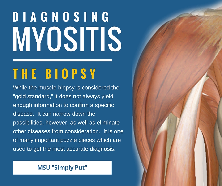
The muscles often selected for sampling are the bicep (upper arm muscle), deltoid (shoulder muscle), or quadriceps (thigh muscle). The procedure requires only a few small pieces of tissue to be removed from the designated muscle. The tissue and cells removed are viewed microscopically. The laboratory report can often take several weeks for results to be read and released. The muscle biopsy is considered the “gold standard” in diagnosing myositis and can rule out other diseases.
It is recommend that muscle biopsies be performed by a surgeon with experience or by a neuromuscular disease specialist. Obtaining adequate specimens and knowing how to properly handle the muscle tissue specimen is important. If not properly performed, the biopsy results will be skewed and the patient may need a repeat biopsy. However, if a surgeon unfamiliar with Myositis must do the biopsy, it is important that they communicate first with a neuromuscular specialist as well as with the lab that will be evaluating the sample prior to the biopsy.
Needle Muscle Biopsy: This procedure is performed by inserting a biopsy needle into the muscle and removing a small piece of muscle from an area suspected of weakness and/or inflammation. The location of the biopsy will be numbed using a topical agent such as Lidocaine. This method is less invasive, but the risk of extracting tissue which has no inflammation exists.
Surgical Muscle Biopsy: Surgical biopsies can be performed under local or general anesthesia, depending on the biopsy site and the surgeon’s preference and is typically considered an outpatient procedure. A small incision is made into the selected site and 4-5 small strips of muscle tissue are extracted. The incision size will vary by location and the surgeon performing the biopsy, but it is typically anywhere from 2-4 inches long.
Skin Biopsy
A small piece of skin is removed for laboratory analysis. A positive skin sample can confirm the diagnosis of Dermatomyositis and rule out other disorders, such as Lupus. If the skin biopsy confirms the diagnosis, a muscle biopsy may be unnecessary.
It is important to note that the skin biopsy for dermatomyositis and Lupus can appear identical under a microscope. Having an experienced dermatologist or rheumatologist is helpful when interpreting the results of the biopsy together with clinical features.
MRI (Magnetic Resonance Imaging)
An MRI gives a cross-section image of muscles in a specific area to show if inflammation exists. Inflammation shows as edema, or swelling, and may show a pattern to help differntiate between polymyositis, dermatomyositis, and inclusion body myositis.
An MRI is often useful in selecting which muscle to biopsy.
Chest X-ray
An x-ray is a simple way to check for symptoms of lung damage or disease. Lung disease can accompany Myositis, so if Myositis is suspected or confirmed, this test is frequently conducted.
Pulmonary Function Tests
Pulmonary function tests are a group of tests that measure how well the lungs take in and release air and how well they move gases such as oxygen from the atmosphere into the body’s circulation. For those with lung involvement, this test may be performed at various intervals to determine improvement or disease progression.
Swallowing Tests
If swallowing muscles are involved, a barium swallow test, and others, may be used. This test can be used to determine the cause of painful swallowing, difficulty with swallowing, abdominal pain, bloodstained vomit, or unexplained weight loss. Barium sulfate is a metallic compound that shows up on X-rays and is used to help see abnormalities in the esophagus and stomach. Learn more about dysphagia.
Cancer screenings
Because of a high cancer risk found in some forms of Myositis, both before and after diagnosis, doctors will often perform cancer screenings at the onset or suspicion of Myositis. Cancer may continue to be a risk, or increased risk, in part due to the immune-suppressing drugs used to treat Myositis, so cancer screenings should be continued on a regular basis. Your doctor may advise more frequent screenings than suggested for the patient’s age group.
New Classification Criteria for Inflammatory Myopathies
Evidence-Based Criteria
(Updated 12/19/2018)
For decades we have been using the “Bohan and Peter criteria,” originally proposed in 1975, for diagnosing myositis. While it serves some patients well and provided needed diagnostic criteria at the time, it is not based on hard science. It has severe limitations in which it does not recognize Inclusion Body Myositis (IBM), Necrotizing Autoimmune Myopathy (NAM), or Amyopathic Dermatomyositis (ADM), and it can lead to errors diagnosing inflammatory vs. non-inflammatory myopathies and muscular dystrophies.
The International Myositis Classification Criteria Project (IMCCP), after many years of hard work, and composed of a large number of prominent myositis physicians and researchers across the globe, developed these evidence-based criteria for both adult and juvenile forms of myositis.
Along with the published criteria, there is also a web-based calculator that can be used to determine the probability of a myositis diagnosis and subtype, both with and without muscle biopsy information.
This is so important in advancing a future for myositis patients and ensuring a quicker diagnosis and better treatments. We thank all who are a part of this effort.
Attempts to re-classify, over the years
Almost a dozen attempts have been made to establish diagnostic and classification criteria for patients with idiopathic inflammatory myopathies (IIMs). View the history here >>
Simply Put
“Simply Put” is a service of Myositis Support and Understanding, to provide overviews of Myositis-related medical and scientific information in understandable language.
MSU volunteers, who have no medical background, read and analyze often-complicated medical information and present it in more simplified terms so that readers have a starting point for further investigation and consultation with healthcare providers. The information provided is not meant to be medical advice of any type.


