Clinically Amyopathic Dermatomyositis (CADM)
Amyopathic Dermatomyositis (ADM) and Hypomyopathic Dermatomyositis (HDM), together referred to as Clinically Amyopathic Dermatomyositis (CADM), are subsets of Dermatomyositis (DM), one of the Idiopathic Inflammatory Myopathies. The cause is unclear and there is no cure.
Like “classic” DM, CADM presents with similar skin manifestations and may be associated with lung disease or cancer, however, there is little-to-no evidence of muscle involvement on exam or testing.
CADM is a distinct clinical entity with unique features and autoantibody profiles different from “classic” dermatomyositis.
CADM is also referred to as skin-predominant dermatomyositis and “dermatomyositis siné myositis.”
Signs and Symptoms of CADM
Symptoms vary from patient to patient. Not all patients display all of these symptoms.
- Skin rashes and discolorations which affect sun-exposed areas, cheeks, nose, shoulders, upper chest, and elbows.
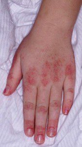
- Gottron’s papules or Gottron’s sign (bumps found over the knuckles, elbows, and knees, often with a raised, scaly break-out)
- Itching and changes in the scalp resembling psoriasis and hair loss
- Ragged cuticles, prominent blood vessels on the nail folds, and other nail-bed abnormalities
- Nonerosive inflammatory polyarthritis
- Joint inflammation
- Lung problems such as Interstitial Lung Disease (ILD), Pulmonary Fibrosis, or Labored breathing (dyspnea)
- Difficulty swallowing (dysphagia)
- Dry, scaly, or rough skin
- Fatigue (may be life-altering and disturb sleep patterns)
- Weight loss
- Low-grade fever
- Muscle pain (myalgia)
Other symptoms and complications
Some patients may develop one or two complications, but rarely all of them.
- Stomach (gastrointestinal) ulcers and intestinal perforations, also more common in children
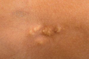
- Calcinosis (hard, often painful lumps or sheets of calcium that form under the skin’s surface)
- Raynaud’s phenomenon
- A hoarse or changing voice (dysphonia)
- Inflammation of the fat which can feel like little bumps lying just under the skin and can be tender to the touch (panniculitis)
- Irregular heartbeat (arrhythmia)
- Inflammation of areas in the heart, myocardium or inflammatory cardiomyopathy
- Other connective tissue diseases (overlaps)
- Cardiovascular disease
- Complications and side effects related to treatment
- Increased risk of having certain types of cancer
- Aspiration pneumonia
Who is affected by CADM?
CADM is a rare disease. A rare disease in the U.S. is defined as a condition affecting fewer than 200,000 people.
Here is some additional information:
- CADM affects women more than men.
- CADM is more common in young white and Asian females.
- CADM typically affects adults between 40-60 years old.
Antisynthetase Syndrome (ASS)
Approximately 90% of those diagnosed with Antisynthetase syndrome experience a form of myositis, either polymyositis or dermatomyositis. This complex syndrome also includes interstitial lung disease (ILD), Raynaud’s phenomenon, inflammatory polyarthritis, mechanic’s hands, and fever.
Mixed Connective Tissue Disease (MCTD)
Mixed connective tissue disease is a rare autoimmune disease considered an “overlap” of three diseases; Systemic Lupus Erythematosus, Scleroderma, and Polymyositis.
Overlap Syndrome
Overlap Syndrome means having more than one autoimmune disease at the same time. This can include many diseases such as Lupus, Dermatomyositis, Polymyositis, Scleroderma, Raynaud’s phenomenon, and Sjögren’s syndrome.
Cancer and CADM
About one-third of DM patients develop malignancy, often within 5 years before or after disease onset; breast and ovarian cancers in women, and lung and prostate cancer in men.
Other less frequently reported cancers include colorectal cancer, non-Hodgkin lymphoma, pancreatic, gastric and bladder cancer.
Cancer can be an underlying cause of dermatomyositis and may precede its onset. If cancer precipitated DM, removal of the cancer may result in remission of dermatomyositis.
Dermatomyositis in adults has also been linked to an increased likelihood of developing cancer post-diagnosis, particularly cancers of the cervix, lungs, pancreas, colorectal system, breasts, ovaries and gastrointestinal tract and non-Hodgkin’s lymphoma. The risk of cancer appears to level off about five years after onset of DM. The incidence of cancer is higher in dermatomyositis than in polymyositis, and particularly in those with certain antibodies. It is strongly suggested that appropriate cancer screenings be conducted upon diagnosis and in the years after a myositis diagnosis.
Because of a high cancer risk, both before and after diagnosis, doctors will often perform cancer screenings at the onset or suspicion of dermatomyositis. Cancer will continue to be a risk with such patients, and cancer screening should be continued on a regular basis and more often than suggested for the patient’s age group per doctor’s advice, particularly because of the increased cancer risk with immunosuppressive treatments.
Dermatomyositis Education Sessions
with Dr. Victoria Werth
Characteristics of CADM
Not everyone diagnosed with CADM will display all, or the same symptoms, and the intensity of the symptoms can vary.
Muscle Involvement
With Amyopathic DM, there are little-to-no muscle findings on exam or on diagnostic testing.
Those with Hypomyopathic DM may show diagnostic muscle findings on EMG, MRI, or elevated muscle enzymes (CK/CPK), usually without marked muscle weakness.
Skin Involvement
The main sign and symptom of CADM, and the reason the term “dermato” is used is skin involvement. CADM may be easier to diagnose compared to other forms of Myositis when patients present with typical skin findings such as Gottron’s Papules, Gottron’s Sign, and the heliotrope rash covering the eyelids and extending onto the face.
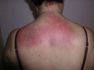
Skin rashes often appear on the eyelids and the face, as well as around the knuckles, nails, knees, elbows, upper back and shoulders (Shawl sign) and chest region (V-sign). Some rashes may be painful, severely itchy (pruritis), and intense enough to disturb sleep.
Scalp inflammation and thinning of the hair is also common.
In rare cases, cutaneous vasculitis, ulcerations, and calcinosis are observed, although more so with juvenile dermatomyositis.
Some commonly used terms:
- Heliotrope refers to the pink-purple-violet shade of the heliotrope flower.
- Violaceous Erythema refers to a violet color rash, often in patches, as a result of injury, irritation, or inflammation causing dilation of the blood capillaries.
- Macular Rash is characterized by a flat red area on the skin that is covered with small confluent bumps (merged together) that may go unnoticed in those with darker skin tones.
- Papules refer to small, raised, solid pimples or swelling, often forming part of a rash on the skin and typically inflamed, but does not produce pus.
Specific Skin Findings of CADM
The appearance of some skin findings are “pathognomonic” for CADM, meaning they are specific to the disease and can help make or confirm a diagnosis. These skin findings can cause major cosmetic issues and are often very itchy. The pattern observed with CADM is the opposite of that found on the hands of patients with Lupus.
- Gottron’s Papules: Red, often scaly, bumps or plaques overlying the knuckles of the fingers, hands, elbows, or knees. Occurs in an estimated 70% of patients.
- Gottron’s Sign: Flat red rash over the back of the fingers, elbows, or knees.
Characteristic and Secondary Skin Findings of CADM
- Shawl Sign: Widespread, flat, reddened rash across the shoulders and upper back, and can extend onto the back of the upper arms and up into the neck and scalp. Pattern of where a shawl would lay if wearing one.
- V-Sign: Widespread, flat, reddened rash covering the chest area, such as where the exposed skin would be when wearing a V-neck shirt.
- Heliotrope Rash: Violaceous (violet or purple) to dusky (can appear as lighter pink, especially in lighter-skin patients) rash over the eyelids, with or without swelling, in a symmetrical (both sides) distribution. In CADM, this rash includes, or hugs, the nasolabial folds (laugh lines) unlike with similar rashes found with Lupus that spare this area.
- Samitz’s Sign: Cuticles can become red and ragged and can be painful. Cuticle overgrowth is also seen in some patients.
- Periungual Telangiectasia: Dilated blood vessels near the nails and under the fingernail.
- Mechanic’s hands: Roughening and cracking of the skin of the tips and sides of the fingers resulting in irregular, dirty-appearing lines that resemble those of a manual laborer.
- Photodistributed Violaceous Erythema: Violet color rash covering the sun-exposed areas such as the face and scalp. This finding shows that the sun can trigger and exacerbate DM/CADM.
- Poikiloderma: A secondary skin finding that includes pigmentary changes, skin atrophy, and telangiectasia (dilated vessels). This is very common with DM/CADM.
- Calcinosis: Often-painful lumps of calcium that can occur in the skin, muscle, and other connective tissues. This is found more with children and adults diagnosed with Juvenile Dermatomyositis (JDM). Surgical intervention may be required for some with severe and painful calcinosis.
- Plugging of the vessels may lead to skin blisters and skin ulcers.
- Scalp Inflammation with thinning of the hair and which may resemble psoriasis.
CADM is a chronic disease with no cure, however, many respond well to certain treatments or combination of treatments with improvement in skin symptoms or even complete remission. As discussed in detail in the Treating Myositis section, finding the most effective treatment for your specific symptoms can be a challenge.
Diagnosing CADM
Which doctors do I see?
CADM is diagnosed and managed by a dermatologist or rheumatologist, usually in coordination with a primary care physician (PCP). Consultation with other specialists may be required depending on your symptoms, other coexisting illnesses, and other organ involvement and may include a cardiologist, pulmonologist, and oncologist.
There are doctors trained in both dermatology and rheumatology which can be especially helpful for those with CADM. See Rheumatologic Dermatology Society (RDS).
When muscle weakness is involved, exercise is important and therefore you may also see a physical therapist, occupational therapist, and if you have trouble swallowing (dysphagia), you may also see a speech-language pathologist.
Clinical and Physical Exam
A complete medical and family history is important in helping to diagnosis a rare and complex disease such as CADM. We suggest writing a detailed medical and family history that you can share with your medical team and that can be used for all future appointments.
Skin findings with CADM can resemble Lupus SLE and are identical on a skin biopsy. An experienced dermatologist can use the skin exam to help better differentiate Lupus from CADM.
Blood Testing
Your doctor will likely order various blood tests when suspecting CADM in order to measure autoimmune and inflammatory markers and to check for myositis-specific and myositis-associated antibodies, and cancer markers, and other antibodies. Some of these tests may include:
- Creatine Kinase (CK or CPK) (muscle enzymes)
- Aldolase
- ALT and AST
- LD (Lactate dehydrogenase)
- Sed Rate (also called ESR or Erythrocyte sedimentation rate)
- ANA (Antinuclear Antibodies panel)
If muscle weakness is evident
EMG and Nerve Conduction Study (NCS)
An EMG (Electromyography) is a test that checks the health of the muscles and the nerves that control the muscles. With dermatomyositis, the EMG may show myopathic changes. EMG testing can help distinguish between weakness due to muscle disease from weakness due to nerve problems. Nerve conduction study is usually done during the same visit and is a test to see how fast electrical signals move through a nerve.
MRI (Magnetic Resonance Imaging)
MRI is being used more frequently for dermatomyositis and can show changes suggesting muscle inflammation. Physicians may also use MRI to choose the best muscle for biopsy.
Muscle Biopsy
The role of a muscle biopsy in diagnosing DM is diminishing as we learn more about myositis antibodies and utilize newly released diagnostic criteria. When certain antibodies or a characteristic skin rash is present, a muscle biopsy may not be necessary.
When a muscle biopsy is necessary, a doctor will remove a small piece of muscle tissue, either via a needle biopsy or an open surgical biopsy, and send it to a lab for testing.
Your doctor will choose the weakest muscle, careful to avoid the site of any recent EMG, for biopsy. An MRI can be helpful in locating the best muscle to biopsy.
Skin Biopsy with skin exam
Biopsying the skin can be helpful in making a diagnosis of CADM. It is vital to find an experienced dermatologist as DM looks identical to Lupus on biopsy and a skin exam can help differentiate the two.
Other Testing
The above tests are not an exhaustive list your physician may use. Other testing will depend on your symptoms and coexisting illnesses. Some examples are below.
Cancer Screening
Since there is an increased risk and potential association of cancer with CADM, your doctor may perform several cancer screenings at diagnosis, and for years following, including those that are age and gender appropriate.
Barium Swallow Test
If you have trouble swallowing, a barium swallow test may be ordered. This is a test often used to determine the cause of painful swallowing, difficulty with swallowing, abdominal pain, bloodstained vomit, or unexplained weight loss. Barium sulfate is a metallic compound that shows up on X-rays and is used to help see abnormalities in the esophagus and stomach.
CT Scan (computed tomography) and Chest X-ray
When lung involvement is suspected, a chest X-ray and CT scan of the lungs can help identify lung disease associated with myositis such as Interstitial Lung Disease (ILD).
Pulmonary Function Tests (PFT’s)
When the lungs are involved, your physician may order PFTs, a group of tests that measure how well your lungs work. This includes how well you are able to breathe and how effective your lungs are able to bring oxygen to the rest of your body.
Challenges of diagnosing CADM
Diagnosing CADM may be a long and difficult process for several reasons.
CADM is a rare disease. Rare diseases are inherently more difficult to diagnose. Do you know the saying, “When you hear hoofbeats, look for horses, not zebras?” This is something doctors are taught and in many cases works well. The “horses” are the most common illnesses and the “zebras” refer to the rare diseases, like CADM.
Each patient presents differently, so a clear pattern or set of symptoms is not necessarily visible to make a diagnosis.
The diagnosis can be delayed when characteristic skin findings are not present.
CADM can mimic other diseases such as systemic lupus erythematosus, pityriasis rubra pilaris, lichen planus, and polymorphous light eruption.
Other involvement with CADM
CADM may affect other organs of the body, cause other illnesses, or place you at a higher risk of these developing.
- Raynaud’s phenomenon. This condition causes your fingers, toes, cheeks, nose and ears to turn pale when exposed to cold temperatures or stress.
- Other connective tissue diseases – Other conditions, such as lupus, rheumatoid arthritis, scleroderma and Sjogren’s syndrome, can occur with CADM (overlap syndromes).
- Cardiovascular complications – CADM can cause heart muscle inflammation (myocarditis). In others who have CADM, heart arrhythmias and congestive heart failure may develop.
- Interstitial Lung Disease (ILD) – Interstitial lung disease refers to a group of disorders that cause scarring (fibrosis) of lung tissue, making the lungs stiff and inelastic.
CADM in photos
Treating CADM
The goals of treatment for CADM are to reduce skin inflammation, reduce morbidity, and improve a patient’s quality of life.
There are no approved therapies for CADM, aside from oral, topical and IV steroids, which are not recommended for long-term use.
Finding a combination of medications may be necessary for successful results.
Sun Avoidance and protective measures
Sun avoidance, ample use of high SPF sunscreen, UVA and physical blockers are recommended. UV protected apparel, large brim hats, gloves, and sunglasses are also very helpful.
Corticosteroids, Topical, Oral, IV
Initially, treatment may consist of topical steroids and non-steroidal topicals. Some may require oral or IV steroids. Corticosteroids address inflammation and simultaneously help suppress the overactive immune system.
Oral and IV steroids have many side effects including weight gain and redistribution of body fat to the face, neck, and abdomen; elevated blood sugars, mood swings, thinning of the skin, cataracts, and osteoporosis.
Additional drugs to suppress the immune system may be added to the steroids depending on disease severity.
Also, depending on disease severity, immunosuppressive medications may be introduced.
Note: Corticosteroids should not be confused with anabolic steroids, used by some weightlifters and athletes, to enhance performance.
Nonsteroidal Topicals
Some of the rashes are treated using nonsteroidal topicals that may include Elidel and topical tacrolimus (Protopic).
Immunosuppressive Drugs
These medications may only be used when muscle involvement is evident.
Steroid-sparing agents, such as immunosuppressive drugs and Disease-Modifying Antirheumatic Drugs (DMARDs), have an added benefit as they often reduce or eliminate the need for steroids while also improving the symptoms of CADM. However, these medications are not without their own risks.
Some commonly used immunosuppressive drugs include azathioprine (Imuran), mycophenolate mofetil (Cellcept), and Tacrolimus (Prograf).
Methotrexate (Rheumatrex) is often used and has been found to help with skin symptoms. It is available in pill form and as an injectable. For those with lung involvement, discuss the risks of using methotrexate.
Cytotoxic Drugs
Cytotoxic medications are a class of immunosuppressives that were originally developed and are still used, to treat certain types of cancer.
Cyclophosphamide (Cytoxan) is one of these medications used. It is available in a pill form and can be given intravenously (IV).
Other Treatments
Antimalarial agents
Hydroxychloroquine (Plaquenil) is one of the commonly used drugs in this class and is used to treat the skin symptoms of CADM. Others used may include chloroquine and quinacrine.
Immunoglobulin Therapy (IVIg, SubQ Ig)
Immunoglobulin comes from antibodies extracted from the plasma from thousands of blood donors. IVIg is the intravenous (IV) method. SubQ Ig is also available, and can be approved for some, and is infused quicker under the skin and eliminates the need for continuous home nursing.
Rituximab (Rituxan)
Rituximab belongs to a class of drugs called monoclonal antibodies. It is given intravenously (IV).
Living with CADM
Day-to-day management of CADM can take many forms. Adapting to a “new normal” can be challenging and require many lifestyle changes.
Exercise Program
It was once thought exercise was not good for myositis patients and caused muscle damage. This has now been proven false through research.
We now know there are massive benefits in having an approved exercise program. This may start with your doctor, physical therapy, or a rehabilitation center. Exercise plans that are supervised by your medical and healthcare team are most effective so you do not injure yourself.
Before starting any type of exercise plan, talk to your doctor first.
Diet and Nutrition
Eating a well-balanced diet is beneficial for those with CADM, and is for your overall health.
Corticosteroids have many side effects including weight gain and redistribution of body fat to the face, neck, and abdomen, elevated blood sugars, and osteoporosis, so you may need to make adjustments in your diet for these conditions.
Ask your doctor about nutrition and a referral to a dietician or nutritionist.
Support
When living with CADM, having a good support system in place is extremely helpful.
CADM is chronic illness and some people in your life may not understand the challenging aspects of the symptoms, treatments, and stress involved in managing this complex, sometimes even invisible illness.
We offer several online support options for patients, family members, caregivers, and friends of those living with CADM. Having an emotional outlet is helpful, and our groups are also educational, resourceful, and even fun.
MSU provides a free, monthly online video support session just for myositis patients. This is a chance to meet others face-to-face, discuss challenges, share tips, and just talk to others who completely understand. View our Events page for upcoming support sessions.
Have you been diagnosed with CADM?
Get the support you deserve. Join the Myositis Support Community.
Safe, private myositis community for patients and caregivers.
CADM Prognosis
The prognosis of CADM depends on many varying factors including patient response to treatment, the age of onset, the severity of disease and its manifestations, the presence of pulmonary or cardiac involvement, and if there is an associated malignancy.
Some CADM patients respond very well to treatment, while others do not respond at all. Finding the right treatment, or combination of treatments can take months to years.
Long-term steroid use may be a major source of morbidity.
CADM In Focus
What does CADM look like?
Patients with CADM will avoid or limit sun exposure as this exacerbates skin symptoms and even causes flares. You may see people wearing UV protected long-sleeved apparel, hats, and gloves, even on the hottest days and during the summer months, to protect themselves as best as possible from the sun.
When the lungs are involved patients may wear oxygen.
Some may look perfectly healthy on the outside, but on the inside, they are sick and struggling on many levels.
Research and Clinical Trials
Research helps us better understand diseases and can lead to advances in diagnosis and treatment. As there are currently no approved treatments for CADM, research is important, as are patients participating in clinical trials. There are several ongoing and upcoming clinical trials for skin-predominant dermatomyositis.
We have partnered with Antidote to help make your clinical trial match easier.
Simply Put
“Simply Put” is a service of Myositis Support and Understanding, to provide overviews of Myositis-related medical and scientific information in understandable language.
MSU volunteers, who have no medical background, read and analyze often-complicated medical information and present it in more simplified terms so that readers have a starting point for further investigation and consultation with healthcare providers. The information provided is not meant to be medical advice of any type.

